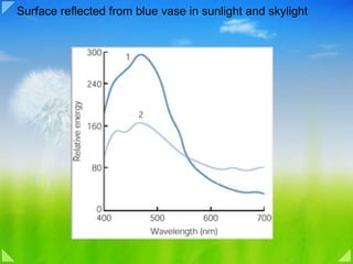
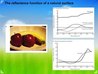
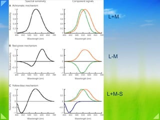
Download to read offline

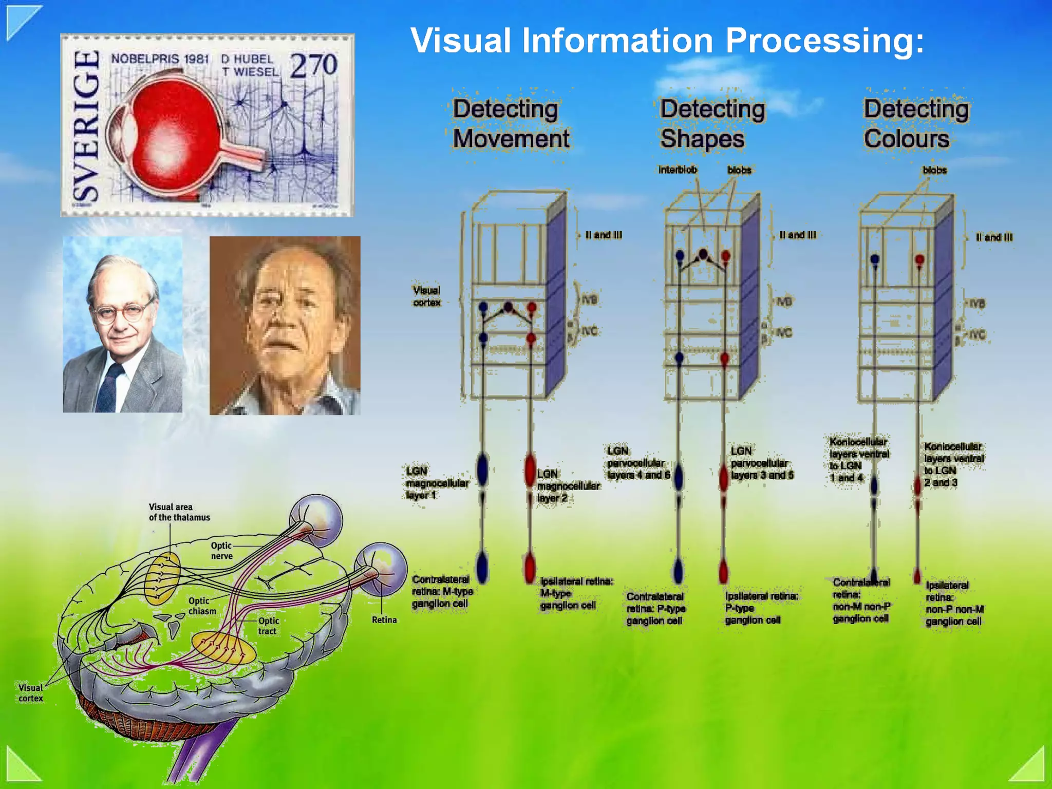
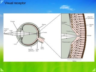
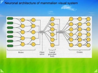
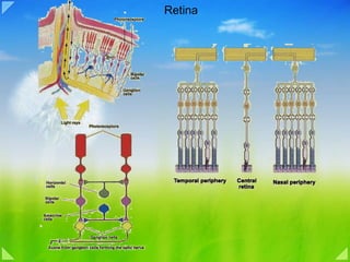
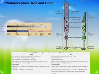
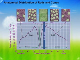
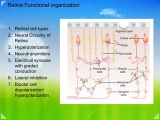
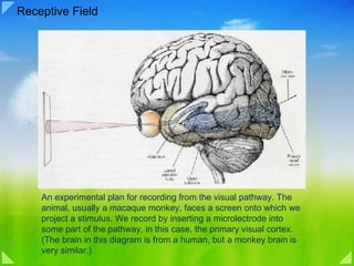
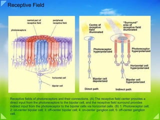
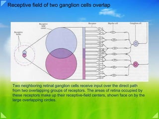
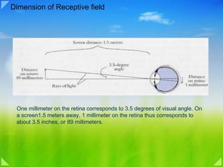
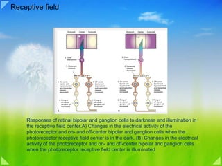
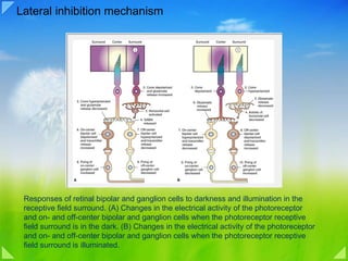
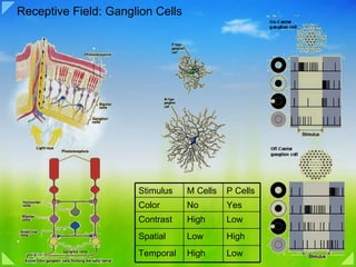
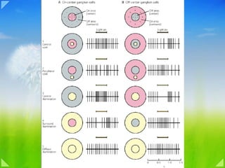
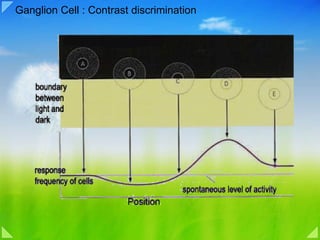
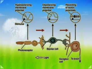
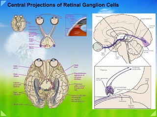
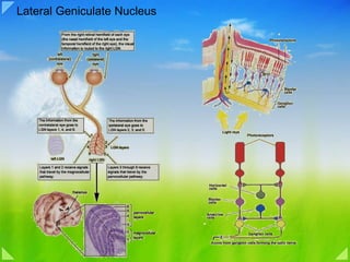
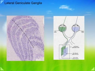
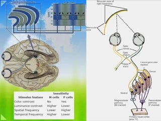
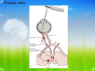
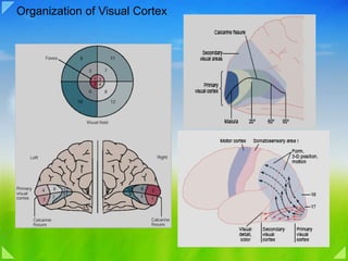
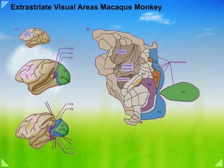
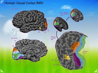

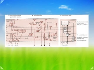
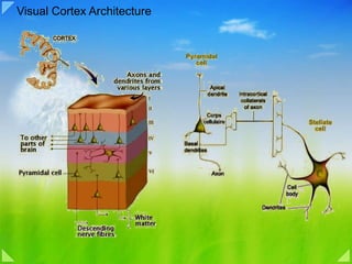
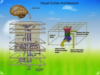
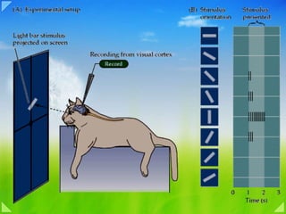
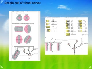
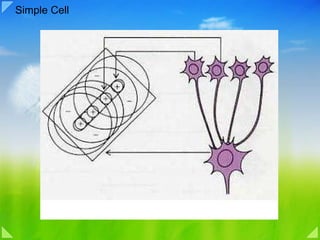
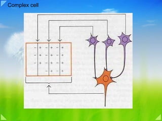
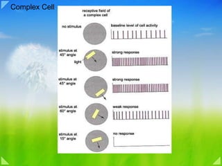

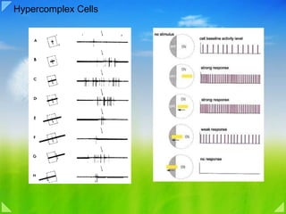
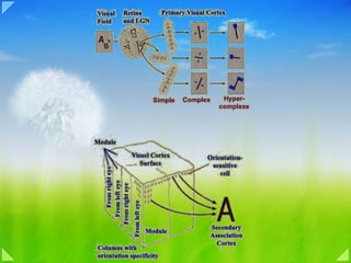
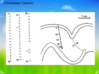
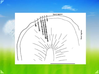
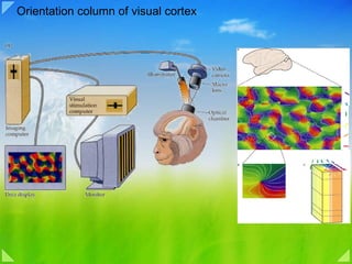

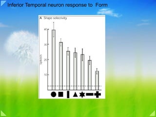
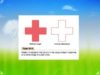
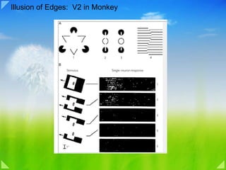
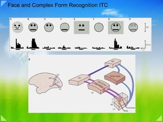

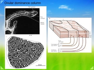
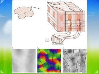
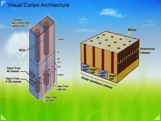
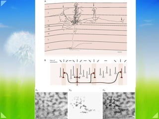

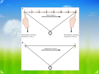
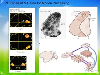
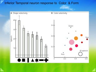

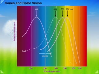
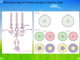


The document discusses the anatomy and physiology of the visual system, including: - The retina contains rod and cone photoreceptors which detect light and have receptive fields. - Ganglion cells in the retina receive input from overlapping photoreceptors and send signals to the lateral geniculate nucleus in the thalamus. - The primary visual cortex, also known as V1, contains cells that respond to features like orientation, motion, and color. Signals from V1 are sent to extrastriate visual areas for further processing.


